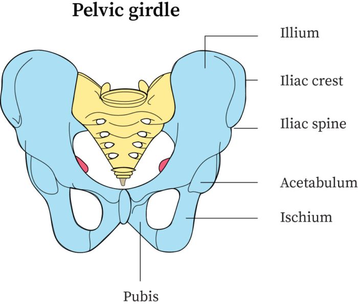
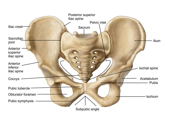
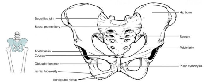
Label the image of the pelvic girdle and delve into the intricate world of human anatomy. This comprehensive guide unravels the structure, functions, and clinical significance of this vital bone structure, providing an immersive exploration into its multifaceted role in supporting the human body.
From its intricate composition to its essential functions, this article delves into the complexities of the pelvic girdle, unraveling its role in movement, stability, and protection.
Pelvic Girdle Anatomy

The pelvic girdle, also known as the pelvis, is a bony structure that forms the lower part of the trunk. It consists of two hip bones (coxal bones), which are joined at the front by the pubic symphysis and at the back by the sacroiliac joints.
Each hip bone is made up of three bones: the ilium, ischium, and pubis.
Bones of the Pelvic Girdle
- Ilium:The ilium is the largest and most superior bone of the pelvic girdle. It forms the upper part of the hip bone and articulates with the sacrum posteriorly and the pubis and ischium anteriorly.
- Ischium:The ischium is located inferior to the ilium and forms the lower and posterior part of the hip bone. It articulates with the ilium superiorly, the pubis anteriorly, and the sacrum posteriorly.
- Pubis:The pubis is located inferior to the ilium and anterior to the ischium. It forms the anterior part of the hip bone and articulates with the ilium superiorly, the ischium posteriorly, and the opposite pubis medially.
Joints of the Pelvic Girdle, Label the image of the pelvic girdle
- Sacroiliac Joint:The sacroiliac joint is a synovial joint that connects the ilium of the hip bone to the sacrum. It is a strong joint that allows for some movement between the pelvis and the spine.
- Pubic Symphysis:The pubic symphysis is a cartilaginous joint that connects the two pubic bones at the front of the pelvis. It allows for some movement between the two hip bones.
Ligaments of the Pelvic Girdle
- Sacroiliac Ligaments:The sacroiliac ligaments are a group of ligaments that connect the ilium of the hip bone to the sacrum. They help to stabilize the sacroiliac joint.
- Pubic Ligaments:The pubic ligaments are a group of ligaments that connect the two pubic bones at the front of the pelvis. They help to stabilize the pubic symphysis.
Functions of the Pelvic Girdle

The pelvic girdle, composed of the hip bones, sacrum, and coccyx, plays a crucial role in supporting the body’s weight, facilitating movement and stability, and protecting internal organs.
Supporting Body Weight
The pelvic girdle is a weight-bearing structure that transmits the weight of the upper body to the lower limbs. The strong, rigid structure of the pelvis helps distribute weight evenly, preventing excessive pressure on any one bone.
Facilitating Movement and Stability
The pelvic girdle provides a stable base for the spine and lower limbs. It allows for a wide range of movements, including flexion, extension, rotation, and abduction of the legs. The pelvic girdle also contributes to balance and coordination during activities like walking, running, and jumping.
Protective Function
The pelvic girdle forms a protective ring around the pelvic cavity, shielding vital organs such as the bladder, rectum, and reproductive organs. The thick, muscular walls of the pelvis provide a barrier against external impacts and injuries.
Clinical Significance of the Pelvic Girdle

The pelvic girdle plays a crucial role in various aspects of human health and well-being. Its importance is particularly evident in childbirth, where it provides a stable and supportive framework for the passage of the baby during delivery.
Childbirth
During childbirth, the pelvic girdle undergoes significant changes to accommodate the passage of the baby’s head and body. The ligaments and muscles of the pelvic girdle relax and stretch, allowing the pelvic bones to widen and the birth canal to enlarge.
This process, known as pelvic dilation, is essential for ensuring a safe and successful delivery.
Injuries and Disorders
The pelvic girdle is susceptible to various injuries and disorders, ranging from minor strains and sprains to more severe fractures and dislocations. Common injuries include:
- Pelvic fractures:These can occur due to high-impact trauma, such as car accidents or falls.
- Pelvic dislocations:These involve the displacement of one or more pelvic bones from their normal position.
- Sacroiliac joint dysfunction:This refers to pain and inflammation in the sacroiliac joint, which connects the sacrum to the pelvis.
- Pubic symphysis diastasis:This is a condition in which the pubic bones separate during pregnancy or childbirth.
Imaging Techniques
Various imaging techniques are used to assess the pelvic girdle and diagnose any underlying injuries or disorders. These include:
- X-rays:These provide a basic visualization of the pelvic bones and can detect fractures or dislocations.
- Computed tomography (CT) scans:These provide more detailed cross-sectional images of the pelvic girdle, allowing for the evaluation of bone structure and soft tissue damage.
- Magnetic resonance imaging (MRIs):These provide highly detailed images of the pelvic girdle, including muscles, ligaments, and nerves, making them useful for diagnosing soft tissue injuries.
Labeling the Image of the Pelvic Girdle: Label The Image Of The Pelvic Girdle
The pelvic girdle, also known as the hip bone, is a complex structure composed of several bones that form the pelvis. It provides support for the lower extremities, protects the pelvic organs, and facilitates childbirth. To accurately identify the individual bones of the pelvic girdle, it is essential to examine a high-quality image and label them accordingly.
The following table provides a detailed description of each bone of the pelvic girdle, along with its location:
| Bone | Description | Location |
|---|---|---|
| Ilium | The largest and most superior bone of the pelvic girdle, forming the upper and lateral portions of the pelvis. | Forms the crest of the ilium and the anterior superior iliac spine. |
| Ischium | The inferior and posterior bone of the pelvic girdle, forming the lower and posterior portions of the pelvis. | Forms the ischial tuberosity and the ischial spine. |
| Pubis | The anterior and inferior bone of the pelvic girdle, forming the anterior and inferior portions of the pelvis. | Forms the pubic symphysis and the pubic tubercle. |
| Sacrum | A triangular bone located posteriorly, connecting the pelvic girdle to the vertebral column. | Forms the sacral promontory and the sacral hiatus. |
| Coccyx | A small, triangular bone located posteriorly, representing the vestigial tailbone. | Forms the apex of the pelvis. |
Refer to the provided high-quality image of the pelvic girdle and label the bones using HTML tags. The labeled image will aid in visualizing the location and relationships between these bones.

Popular Questions
What is the pelvic girdle?
The pelvic girdle is a ring-shaped structure formed by the hip bones (ilium, ischium, and pubis) that connects the lower limbs to the axial skeleton.
What are the functions of the pelvic girdle?
The pelvic girdle provides support for the body’s weight, facilitates movement and stability, and protects internal organs.
What are some common injuries or disorders affecting the pelvic girdle?
Common injuries or disorders affecting the pelvic girdle include fractures, dislocations, and arthritis.
What imaging techniques are used to assess the pelvic girdle?
Imaging techniques used to assess the pelvic girdle include X-rays, CT scans, and MRIs.
